Macula
The macula, an area of the retina of humans and other old-world primates, is centered on the fovea, the area of highest acuity vision.
The 6-mm diameter human macula is defined by neurobiologists as the area where ganglion cells form a continuous layer, by ophthalmologists as the area inside the arcades formed by the retinal circulation, and by epidemiologists as the area inside the grading grid of the Early Treatment of Diabetic Retinopathy Study and the Wisconsin Age-related Maculopathy Grading System. [1-4]
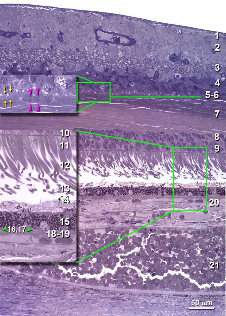
The macula comprises numerous clinically relevant layers, sublayers, and potential spaces.
Retinal layers (from inner to outer)
- Nerve fiber layer (NFL; 1)
- Ganglion cell layer (GCL; 2)
- Inner plexiform layer (IPL; 3)
- Inner nuclear layer (INL; 4)
- Outer plexiform layer (OPL; 5,6,7) in macula has sublayers of
- Bipolar and horizontal cell dendrites
- Cone pedicles and rod spherules
- Henle fibers (photoreceptor inner fibers and Müller cells).
- Outer nuclear layer (ONL; 8,9,10)
- Rod nuclei (8)
- Cone nuclei (9)
- Outer fibers (10; in fovea only; not seen here)
- Photoreceptors (Polyak’s bacillary layer)
- Inner segments (IS; 11,12)
- Myoid (11)
- Ellipsoid (12)
- Outer segments (OS; 13)
- Subretinal space (14)
- Retinal pigment epithelium (RPE; 15)
- Basal lamina of the RPE (16)
- Sub-RPE space (17)
Choroidal layers
- Bruch’s membrane (BrM)
- Inner BrM (18)
- Outer BrM (19)
- Choriocapillaris (20)
- Choroid, delimited from the sclera by the lamina suprachoroidea.
Stereotypic extracellular deposits accumulate with aging and age-related macular degeneration (AMD)
- Subretinal space (subretinal drusenoid deposit; 14)
- Internal to the RPE basal lamina (basal laminar deposit; BlamD; 16); containing basal mounds (Bmound)
- Sub-RPE space (between the RPE basal lamina and Bruch’s membrane, BlinD/drusen; 17)

Aging and AMD macula, revised from Reference 5; Panel A: 1, RPE basal lamina; 2, inner collagenous layer; 3, elastic layer; 4, outer collagenous layer; 5 choriocapillary endothelium basal lamina; Panel B, see main text.
References
- Polyak SL: The Retina. Edited by Chicago, University of Chicago, 1941,
- Hogan MJ, Alvarado JA, Weddell JE: Histology of the Human Eye. An Atlas and Textbook. Edited by Philadelphia, W. B. Saunders, 1971, p.pp. 328-363
- Klein R, Davis MD, Magli YL, Segal P, Klein BEK, Hubbard L: The Wisconsin Age-Related Maculopathy Grading System, Ophthalmol 1991, 98:1128-1134 PMID 1843453
- Curcio CA, Messinger JD, Mitra AM, Sloan KR, McGwin G, Jr, Spaide R: Human chorioretinal layer thicknesses measured using macula-wide high resolution histological sections, Invest Ophthalmol Vis Sci 2011, 52:3943-3954 PMID 21421869
- Curcio CA, Johnson M, 2012. Structure, function, and pathology of Bruch’s membrane. In Retina, S.J. Ryan, A.P. Schachat, C.P. Wilkinson, D.R. Hinton, S. Sadda, P. Wiedemann, eds. (London, Elsevier).

